What is Optogenetics?

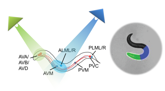
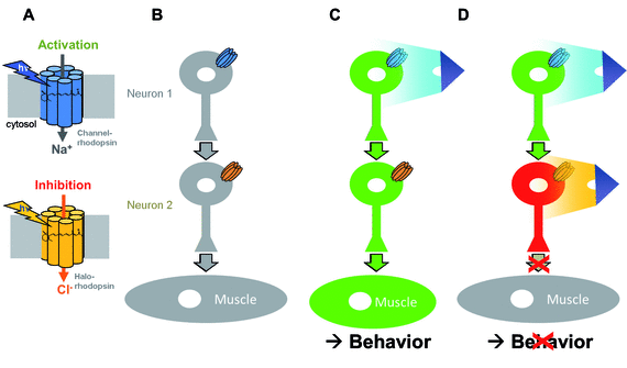
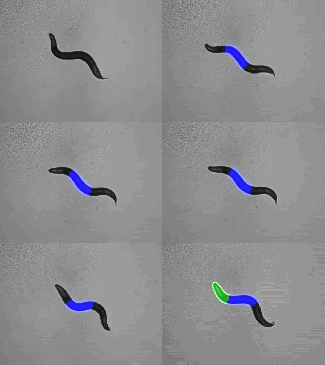
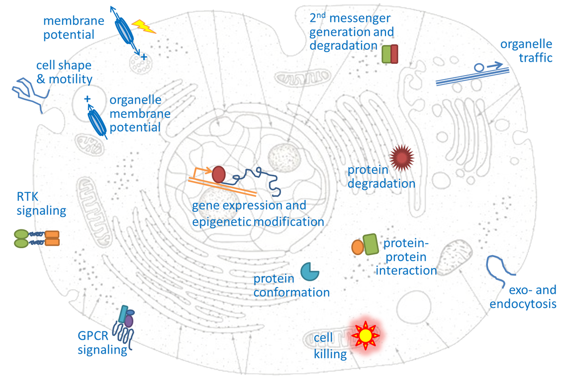
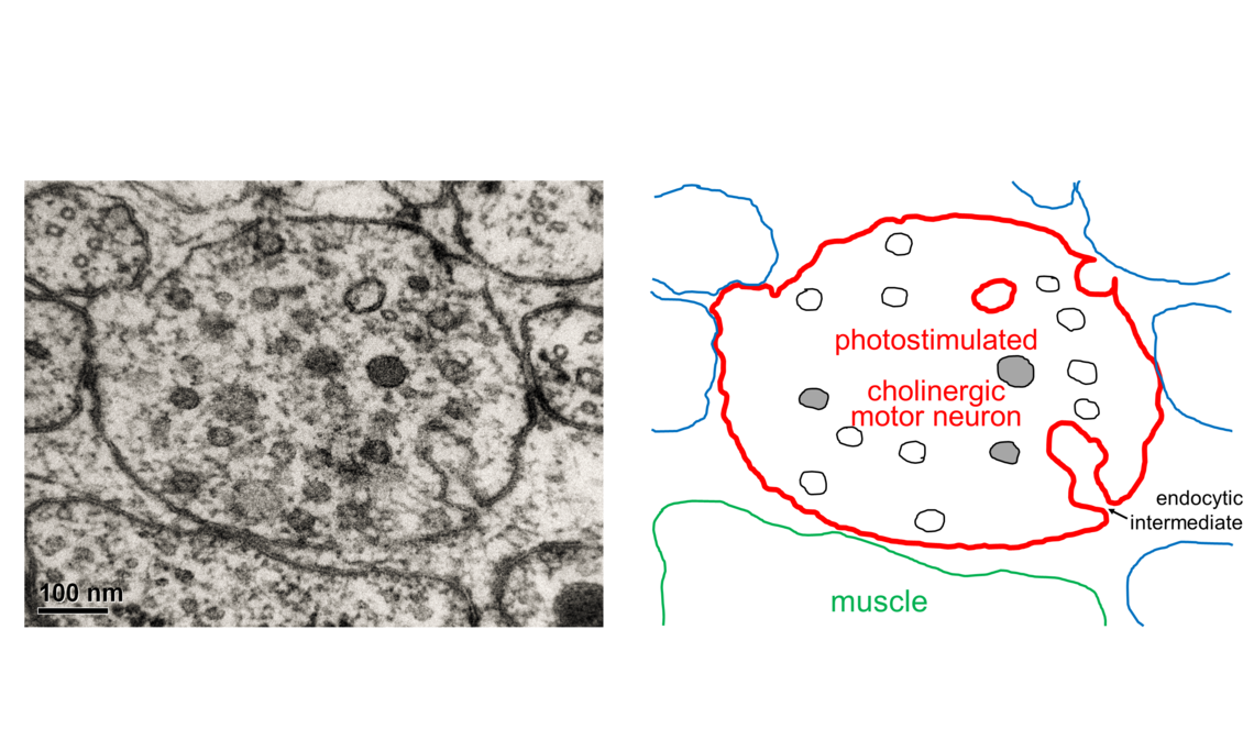
“Optogenetics” summarizes methodologies and applications that utilize genetically encoded (or addressable) light-controlled proteins or molecules, which enable temporally precise control of molecular, cellular or (neuronal) network activities in live cells, tissue and animals. Optogenetic actuators are proteins found in natural light-sensitive organisms (often, these are microbes), whose genetic code is transfered to cells of higher animals, for example laboratory 'model' animals like nematodes, flies, fish and mice. These actuators, for example the famous channelrhodopsins, enable manipulating ion fluxes and membrane voltage. Upon illumination, these proteins open an ion-selective pore, thus leading to altered membrane voltage within milliseconds, and thereby, in the case of neurons or (cardiac) muscle cells, activating the cell. The consequence is precisely triggered neuronal firing, or muscle twitching, for example leading to locomotion of the animal or a heart beat. An optogenetic 'counterplayer' of channelrhodopsin is halorhodopsin, that pumps chloride ions into cells, thus inhibiting their activity. Other optogenetic actuators generate so-called 2nd messengers, which modify cellular signaling or the biochemistry of the cell. Further optogenetic tools were developed that allow the researcher to control gene expression, protein interaction or degradation, or cell death. Apart from natural or engineered light-sensitive proteins, optogenetic tools can be proteins that are made photoswitchable by small chemical compounds, which are often linked physically to the protein of interest. Last, optogenetics also encompasses genetically encoded optical sensors for Ca2+, membrane voltage, pH, small molecules, or 2nd messengers.

Historical Perspective

First ‘optogenetic’ experiments were performed in the lab of Gero Miesenböck (University of Oxford), who transferred the photosensory protein cascade from the eye of the fruitfly Drosophila to mammalian neurons and could evoke activity upon light application (Zemelman et al. 2002). These experiments, while somewhat complicated and too slow to trigger neurons at meaningful timescales, sparked an interest in similar approaches. Around the same time, a ‘photopharmacological’ approach was promoted by Isacoff, Trauner and Kramer: They first used untethered inhibitors of ion channels, which were photoswitchable from inactive to active conformations, in order to trigger neuronal activity; later these chemical photoswitches were tethered to modified channels, thereby achieving genetic addressability (Banghart et al. 2004; Szobota et al. 2007). The most successful and widely used optogenetic tools, i.e. the microbial rhodopsins, have their origins in Germany. In 2002 and 2003, Nagel, Hegemann and Bamberg described the function of channelrhodopsins 1 and 2 (ChR1 and ChR2) as directly light-gated ion channels (Nagel et al. 2002; Nagel et al. 2003). In the 2002 paper, they stated: “Moreover, the ability of ChR1 to mediate a large light-switched H+ conductance in oocytes holds promise for the use of ChR1 as a tool for measuring and/or manipulating electrical and proton gradients across cell membranes, simply by illumination.” In 2003: “Additionally, we have shown that expression of ChR2 in oocytes or mammalian cells may be used as a powerful tool to increase cytoplasmic Ca2+ concentration or to depolarize the cell membrane, simply by illumination.” These two papers raised the interest of a number of groups (Deisseroth, Gottschalk, Herlitze, Pan, Yawo) who implemented expression of these proteins in excitable cells, i.e. muscles, neurons and nervous systems, to activate these cells by light, and even to evoke coordinated behavior (Boyden et al. 2005; Li et al. 2005; Nagel et al. 2005; Bi et al. 2006; Ishizuka et al. 2006). The term ‘optogenetics’ was coined to denote this fundamentally novel and evidently powerful approach (Deisseroth et al. 2006). In 2007, halorhodopsin (NpHR) was introduced as a complementary optogenetic tool to hyperpolarize cells, and its combined use with ChR2 for independent push-pull control over membrane potential, action potential firing, and even coordinated animal behavior was demonstrated (Han and Boyden 2007; Zhang et al. 2007). Subsequently, rhodopsin-based optogenetic tools have been widely used. In recent years, many other optogenetic tools and applications were introduced not only for neuro-, but also for cell biology.

Examples of optogenetic tools and applications


Optogenetic tools do not only address membrane currents and membrane potential, like the rhodopsins, or modifed ion channels in neurons. Tools have been designed by this exploding research field that address also intracellular events and mechanisms. This can involve the expression of genes in the nucleus, the interaction and transport of proteins inside the cell, changing their shape and conformation, so they can interact with other proteins, but also the degradation of proteins, up to indcution of cell death. Also means of signaling at the cell surface, or inside the cell, have been made light sensitive, like 2nd messenger systems, G protein coupled receptors, or receptor tyrosine kinases. Last, shape and mobility of cells, as well as membrane traffic can be addressed using optogenetic tools.

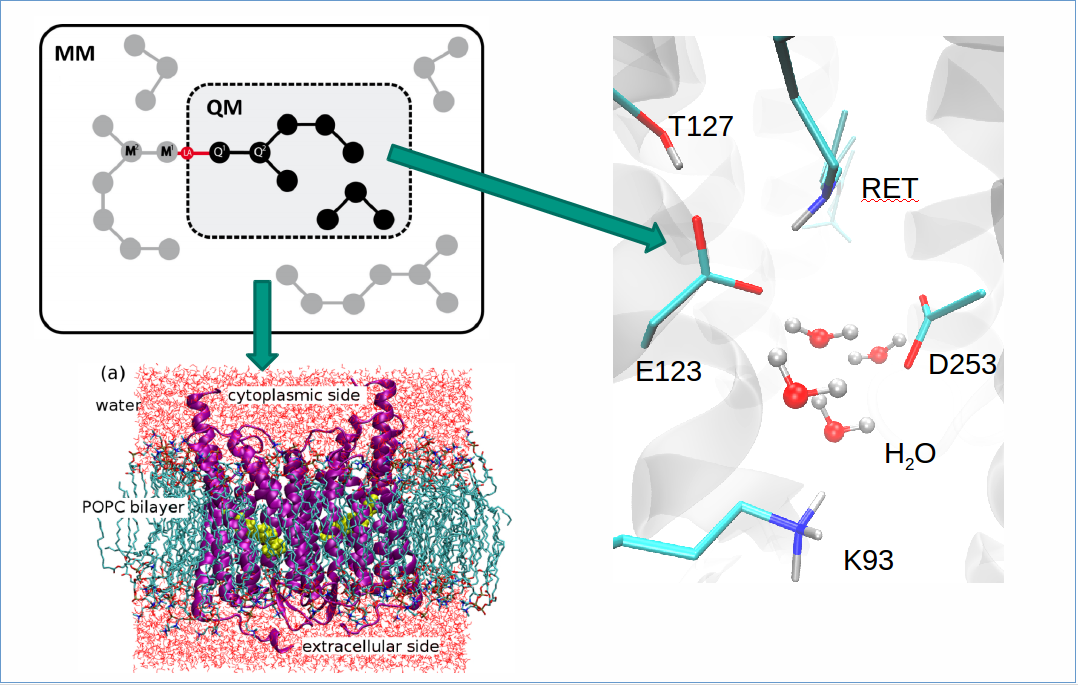
An important aspect of research in optogenetics is to achieve a mechanistic and structural understanding of light-sensitive proteins, such that they can be designed further for improved or highly specific optogenetic applicability.


Optogenetic applications in human therapy?

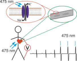
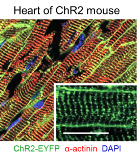
Optogenetic tools also hold great promise for applications in therapy of human diseases. This appears possible for applications in the eye, in degenerative diseases of the retina, or the inner ear, where photoreceptor or sound-sensitive mechanoreceptor cells vanish. However, since the neurons that normally ‘transport’ information from these receptors to the brain are still existent despite degeneration of the receptors, these neurons can be rendered light sensitive by optogenetics. They would respond to (amplified) ambient light (in the eye), or to sound picked up by a microphone that is transferred into light-signals and delivered to the inner ear by specific hardware (for example, arrays of µLEDs). Other applications address the heart, where optogenetic pacemakers, or defibrillation, are being explored. Moreover, there is hope to use optogenetic intervention also in neurodegenerativ or neuropsychiatric disorders, such as Parkinson’s disease, epilepsy, or depression, where particular cell types or brain ‘nuclei’ loose function or evoke uncontrolled activity. Cell-type specific expression of optogenetic actuators promises more selective control than is currently available through electric deep brain stimulation (DBS) or epilepsy surgery. However, it is clear that biomedical applications need a lot more ground work in suitable animal models, accompanied by a public discussion (because of the ethical considerations applying when human neurons are made light-sensitive), before their translation into the clinic.

Ethical considerations

Any modification of the human genome, or the expression of genes in specific cell types, raises concerns and has strong ethical implications. This is also true for the potential use of optogenetic tools in human therapy, as it involves the introduction of foreign genetic material into cells of the patient. Optogenetic applications in the brain require a particularly careful consideration, as there is the fear (and, however, very remote potential) of exogenous induction or modulation of brain activity, and possibly even behavior, that is against the free will of the patient. Thus, principles of ethics in medicine need to cover the novel questions raised by the development and potential use of optogenetics in humans. These principles must then be strictly applied in any possible optogenetic application in humans. We would like to contribute to this discussion and to inform the public about the possibilities, benefits, and risks of optogenetics in humans.